
Ultrasound imaging has become a vital diagnostic tool in veterinary medicine, offering a non-invasive and pain-free way to visualize internal organs and tissues in pets. Similar to its use in human medicine, veterinary ultrasound provides valuable insights into a pet’s health, enabling veterinarians to accurately diagnose and treat a range of conditions, from gastrointestinal issues to cardiovascular disease.
By harnessing the power of high-frequency sound waves, ultrasound technology offered at the pet clinic in Murrieta, CA produces detailed images of internal structures, facilitating timely interventions and improving treatment outcomes for companion animals.
What is veterinary ultrasound?
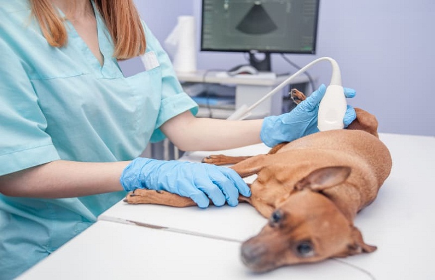
Veterinary ultrasound uses high-frequency sound waves to produce images of internal structures. A probe, or transducer, is placed on the pet’s body, emitting sound waves that bounce off tissues and organs. These echoes are then converted into images on a screen, allowing veterinarians to examine internal anatomy.
Types of ultrasound exams
- Basic Ultrasound: Evaluates general health.
- Targeted Ultrasound: Focuses on specific organs or systems.
- Contrast-Enhanced Ultrasound: Uses specialized agents to highlight structures.
- Doppler Ultrasound: Measures blood flow.
When is veterinary ultrasound applicable?
Ultrasound scans are used in veterinary medicine in the following situations:
- Abdominal Imaging: Evaluates liver, kidneys, spleen, pancreas, and gastrointestinal tract.
- Cardiac Imaging: Assesses heart structure and function.
- Musculoskeletal Imaging: Examines joints, tendons, and ligaments.
- Reproductive Imaging: Evaluates reproductive organs and detects pregnancy.
- Oncology: Monitors tumor growth and response to treatment.
- Foreign Body Detection: Identifies ingested or embedded objects.
- Guided Procedures: Facilitates biopsies, fluid drainage, and catheter placement.
What are the benefits of ultrasound for pets?
The benefits of ultrasound for pets are as follows:
Diagnostic Benefits
- Accurate diagnosis of internal injuries or diseases
- Early detection of conditions, such as cancer or heart disease
- Identification of foreign objects or blockages
- Evaluation of organ function and structure
- Detection of infections or abscesses
Therapeutic Benefits
- Guided biopsies or fluid drainage
- Monitoring of treatment response
- Identification of optimal treatment plans
- Reduced need for exploratory surgery
- Enhanced patient care and management
Convenience and Comfort Benefits
- Non-invasive and pain-free procedure
- No radiation exposure
- Minimal restraint required
- Quick procedure time (usually 15-60 minutes)
- Can be performed on awake or sedated pets
Cost-Effective Benefits
- Reduces the need for additional testing (e.g., CT scans, MRI)
- Minimizes surgical costs
- Early detection can prevent costly complications
- Enhances treatment outcomes, reducing long-term costs
- Cost-effective alternative to exploratory surgery
Emotional Benefits
- Provides peace of mind for pet owners
- Enhances veterinarian-client communication
- Facilitates informed decision-making
- Supports bond between pet owner and veterinarian
- Offers reassurance and emotional support during diagnosis and treatment
Other Benefits
- Portable and accessible technology
- Digital image storage for record-keeping
- Enhanced patient safety
- Supports research and education in veterinary medicine
- Continuously evolving technology for improved diagnostics
How is an ultrasound scan performed for pets?
Here’s a step-by-step guide to the ultrasound procedure for pets:
Pre-Procedural Preparation
- Fasting: Recommended for abdominal ultrasounds (6-12 hours)
- Withdrawal of food and water: To prevent gastrointestinal interference
- Sedation or anesthesia: May be necessary for anxious or uncooperative pets
- Shaving: May be required for optimal image quality
- Cleaning: Skin cleaned with an antiseptic solutio
Procedure Steps
- Positioning: Pet placed in a comfortable position (e.g., lateral recumbency)
- Probe application: Transducer applied to skin with ultrasound gel
- Image acquisition: Veterinarian captures images of internal structures
- Scanning: The probe moved systematically to examine organs and tissues
- Measurement and annotation: Images measured and annotated for future reference
What are the advances in veterinary ultrasound?
Veterinary medicine has evolved tremendously over the recent decades with the following advances in ultrasound scans:
- Portable Units: Enhance accessibility.
- Digital Image Storage: Facilitates record-keeping.
- 3D and 4D Imaging: Provides enhanced visualization.
- Artificial Intelligence: Aids in image analysis.
Veterinary ultrasound has transformed the diagnostic landscape, offering a safe, effective, and informative tool for pet owners and veterinarians. By understanding the applications, benefits, and limitations of ultrasound imaging, pet owners can make informed decisions about their pet’s healthcare.